Model Introduction
Osteoporosis is a metabolic bone disease characterized by reduced bone mass and deterioration of bone tissue microarchitecture, leading to decreased bone strength and an increased risk of fractures. Based on etiology, it can be classified into primary (e.g., postmenopausal osteoporosis, senile osteoporosis) and secondary osteoporosis (e.g., glucocorticoid-induced osteoporosis, disuse osteoporosis, etc.).
Currently, commonly used animal models for osteoporosis include: ovariectomy (OVX) models, drug-induced models, nutritional models, disuse models, brain-derived models, genetic models, and combined models. Different models simulate various pathogenic mechanisms, providing experimental platforms for mechanism research and intervention strategy development.
Research Applications
Osteoporosis animal models can be used to:
- Simulate bone loss caused by estrogen deficiency post-menopause.
- Study glucocorticoid-associated secondary osteoporosis.
- Analyze the impact of nutritional deficiencies on bone mineral density.
- Study bone metabolism abnormalities caused by long-term immobilization or weightlessness.
- Explore the regulation of bone remodeling by the neuroendocrine system.
- Evaluate anti-osteoporotic drugs and intervention measures. Different models have different mechanistic focuses and should be selected based on research objectives.
Experimental Design Key Points
I. Castration/Ovariectomy Methods
Includes surgical castration and chemical castration. 1. Surgical Ovariectomy (OVX)
- Advantages: Single modeling factor, stable model, good reproducibility, simulates bone metabolism characteristics post-menopause.
- Limitations: Sudden drop in estrogen levels in animals differs from the gradual decline in menopausal women.
2. Chemical Castration
- Method: Bone mass loss is caused by inhibiting estrogen secretion.
- Common Drugs: LHRH receptor agonists, GnRH agonists, estrogen receptor antagonists, non-steroidal androgen antagonists, aromatase inhibitors, etc.
- Advantages: Avoids interference from surgical trauma.
- Limitations: Adverse drug reactions and drug interactions may affect experimental results.
II. Drug-Induced Model (Glucocorticoid Model)
Glucocorticoid-induced osteoporosis is a common type of secondary osteoporosis.
- Common Animals: Rats are most commonly used; rabbits and sheep can also be used.
- Key Factors: Animal age, drug dosage, duration of administration.
- Characteristics: Significant bone loss; bone loss is reversible after drug withdrawal.
- Risks: High doses can cause osteonecrosis, immunosuppression, or even death. Animal Characteristics:
- Rats: Good reproducibility, low cost, but lack Haversian systems, have a short bone remodeling cycle, and skeletons do not fully mature.
- Rabbits: Possess complete Haversian systems, fast bone turnover, skeletons can fully mature, but lack sufficient cancellous bone.
- Sheep: Large body size, sufficient sampling material, but modeling is time-consuming and expensive.
III. Nutritional Osteoporosis Model
- Method: Restrict intake of calcium, Vitamin D, protein, etc.
- Significance: Used to study osteoporosis caused by nutritional deficiency.
- Limitations: Complex feed formulas, many confounding factors, time-consuming as a standalone model with low success rates; often used as a supplementary method.
IV. Disuse Osteoporosis Model
- Principle: Limited exercise or functional impairment leads to bone loss.
- Common Methods: Mechanical fixation, tail suspension, tenotomy, sciatic neurectomy, etc.
- Application Scenarios: Long-term bed rest, paralysis, weightlessness (e.g., astronauts). Tail suspension experiments show reduced new bone formation and increased or unchanged bone resorption; significant bone loss occurs after two weeks of suspension.
V. Brain-Derived Osteoporosis Model
- Principle: Destruction of the hypothalamic arcuate nucleus causes endocrine dysfunction, leading to osteoporosis.
- Characteristics: Highly operable, stable, and reproducible.
- Suitable for: Research on the pathogenesis, prevention, and treatment of osteoporosis.
VI. Genetic Modeling Method
Includes transgenic and gene mutation modeling.
- Common Genes: α, β estrogen receptors, aromatase, etc.
- Advantages: Stable systemic osteoporosis manifestations, unaffected by external interference.
- Ideal for: Primary osteoporosis modeling.
VII. Combined Modeling Method
- OVX + Glucocorticoids
- OVX + Low calcium diet
- OVX + Low calcium diet + Glucocorticoids (Triple method yields more pronounced effects)
- Advantages: Shortens modeling time.
- Limitations: Complex interference factors may affect research results.
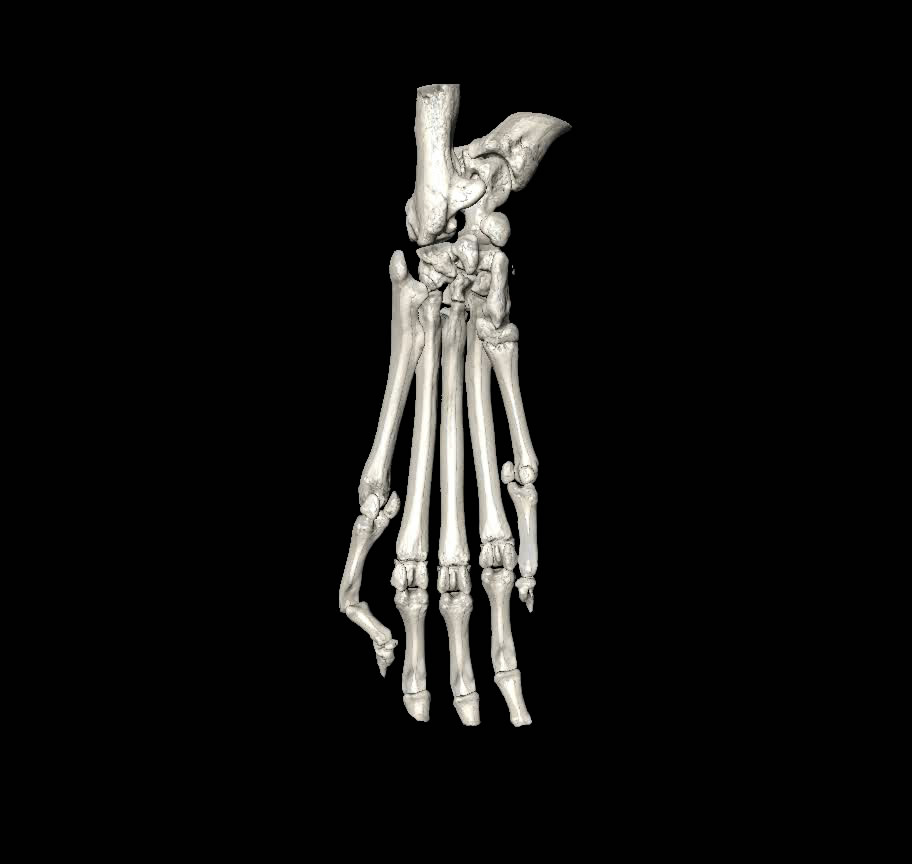
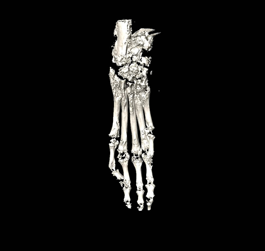
Key Detection Indicators
Evaluation indicators for osteoporosis models are generally consistent and include:
- Bone Mineral Density (BMD) measurement.
- Bone histomorphometry analysis.
- Biochemical marker detection.
- Bone biomechanical testing.
- Body weight changes.
- Gonadal morphology observation. The primary indicators are:
- Changes in bone mass.
- Histopathological changes in bone tissue. Single indicators have limitations; comprehensive evaluation should be conducted from “macro to micro, 2D to 3D, and qualitative to quantitative” to improve model evaluation accuracy.